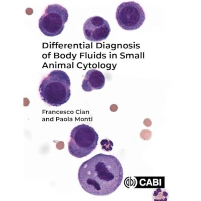
Differential Diagnosis of Body Fluids in Small Animal Cytology
 Beschrijving
Bol
This new volume provides a comprehensive coverage of cytology of all body fluids encountered in dogs and cats. It includes separate chapters for each fluid, from cavitary effusions (pleural, pericardial, and peritoneal) to synovial fluids, tracheal and bronchoalveolar lavages (TWs, BALs), cerebrospinal fluids (CSFs) and urines. Illustrated with high quality photomicrographs, Differential Diagnosis of Body Fluids in Small Animal Cytology provides a comprehensive review of fluid cytology, with an extensive visual atlas. With key points describing the main clinical and cytological features of each pathologic condition, the book provides lists of causes and differential diagnoses, including handy 'pearls and pitfalls' boxes. It is also enriched by chapters on microbiology testing of body fluids and other advanced diagnostic techniques, making the book a valuable resource for veterinary specialists (in particular clinical and anatomical pathologists), residents, veterinary undergraduate students, and small animal practitioners. Key features · Over 180 high-quality photomicrographs. · Ideal reference book with concise descriptions of each pathologic process. · Organised into key bullet points to facilitate use during diagnostic work, or as a revision aid. Illustrated with high quality photomicrographs, Differential Diagnosis of Body Fluids in Small Animal Cytology provides a comprehensive review of fluid cytology, with an extensive visual atlas. With key points describing the main clinical and cytological features of each pathologic condition, the book provides lists of causes and differential diagnoses, including handy 'pearls and pitfalls' boxes. It is also enriched by chapters on microbiology testing of body fluids and other advanced diagnostic techniques, making the book a valuable resource for veterinary specialists (in particular clinical and anatomical pathologists), residents, veterinary undergraduate students, and small animal practitioners. Key features Over 180 high-quality photomicrographs. Ideal reference book with concise descriptions of each pathologic process. Organised into key bullet points to facilitate use during diagnostic work, or as a revision aid.
This new volume provides a comprehensive coverage of cytology of all body fluids encountered in dogs and cats. It includes separate chapters for each fluid, from cavitary effusions (pleural, pericardial, and peritoneal) to synovial fluids, tracheal and bronchoalveolar lavages (TWs, BALs), cerebrospinal fluids (CSFs) and urines. Illustrated with high quality photomicrographs, Differential Diagnosis of Body Fluids in Small Animal Cytology provides a comprehensive review of fluid cytology, with an extensive visual atlas. With key points describing the main clinical and cytological features of each pathologic condition, the book provides lists of causes and differential diagnoses, including handy 'pearls and pitfalls' boxes. It is also enriched by chapters on microbiology testing of body fluids and other advanced diagnostic techniques, making the book a valuable resource for veterinary specialists (in particular clinical and anatomical pathologists), residents, veterinary undergraduate students, and small animal practitioners. Key features · Over 180 high-quality photomicrographs. · Ideal reference book with concise descriptions of each pathologic process. · Organised into key bullet points to facilitate use during diagnostic work, or as a revision aid. Illustrated with high quality photomicrographs, Differential Diagnosis of Body Fluids in Small Animal Cytology provides a comprehensive review of fluid cytology, with an extensive visual atlas. With key points describing the main clinical and cytological features of each pathologic condition, the book provides lists of causes and differential diagnoses, including handy 'pearls and pitfalls' boxes. It is also enriched by chapters on microbiology testing of body fluids and other advanced diagnostic techniques, making the book a valuable resource for veterinary specialists (in particular clinical and anatomical pathologists), residents, veterinary undergraduate students, and small animal practitioners. Key features Over 180 high-quality photomicrographs. Ideal reference book with concise descriptions of each pathologic process. Organised into key bullet points to facilitate use during diagnostic work, or as a revision aid.
AmazonPages: 360, Paperback, Cab International
 Prijshistorie
Prijzen voor het laatst bijgewerkt op:


 Productspecificaties
 Gerelateerde
  Naar shop


